Hip dysplasia
Technique for HD radiographs
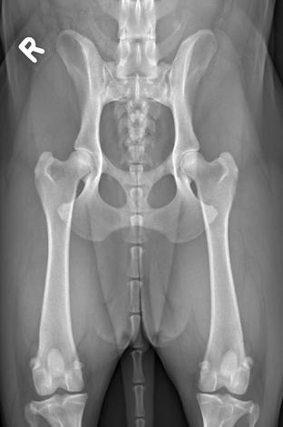
The hip joints can only be evaluated objectively if the pelvis is positioned accurately in the ventro-dorsal view and if the femurs are parallel. To achieve this, the dog must be deeply sedated or anesthetized and placed in a positioning device shaped like a shell. The exposure (kV) should be chosen to sufficiently penetrate the femoral head so that the contour of the acetabulum is clearly visible.
Projections
HD radiograph, extended
The left or right side is identified by a corresponding lead letter. The central beam is directed at the tubera ischiadica. The hind limbs are held at the tarsus and rotated inward until the knee joints are positioned vertically upward. Then, the hind limbs are extended backward until the femurs are as parallel as possible to the X-ray table.
The radiograph should be checked according to the following criteria:
- The pelvis is fully represented, and the patellae must be visible.
- Both obturator foramina appear of equal size and oval shape.
- Both wings of the ilium appear symmetrical.
- The femurs are parallel to each other, parallel to the spine, and as parallel as possible to the X-ray table.
- The patellae are projected between the two femoral condyles.
- The dorsal edge of the acetabulum is visible through the femoral head (otherwise, the image is underexposed).

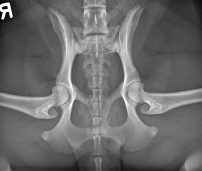
HD radiograph, flexed
The left or right side is identified by a corresponding lead letter. The pelvis is centered, focusing on the hip joints. The hip joints are located at the level of the pectineus muscle, which can be palpated. The knees are laterally spread and flexed. The tarsi are clearly elevated from the table (30-40 cm!) and brought behind the pelvis.
The radiograph should be checked according to the following criteria:
- The seventh lumbar vertebra is also visible, allowing for evaluation of the lumbosacral junction.
- Both obturator foramina appear of equal size.
- The angle between the spine and the femurs is slightly less than 90°.
- The greater trochanter is visible without superposition behind the femoral neck.
- The anterior border of the femoral head-neck junction is located outside the acetabulum.

Images of insufficient quality will not be evaluated. Images of dogs that have not reached the minimum age required for their breed will be provisionally evaluated.

